Examinations in children

Ultrasound
This technique uses ultrasound, the same as that used during pregnancy ultrasounds. It is used to obtain fairly accurate images of the regions scanned. This examination is painless.
Magnetic resonance imaging or MRI
This painless technique produces cross-sectional images of the body. The technique uses the interaction between the natural magnetism of the human body and that of the machine which contains a large magnet. This technique does not involve X-ray irradiation. A contrast product, gadolinium, is sometimes used.
PET Scan
Cancerous cells consume energy so they can multiple in a rapid and unchecked way. This examination is used to identify areas of intense metabolic activity.
First of all, a very small amount of radioactive product is injected: an isotope that binds to active abnormal areas found throughout the body: the primary tumor and or its metastases. The child is then passed through a machine equipped with different sensors that record the radioactivity of the injected substance. A computer then reconstructs the images. The examination is sometimes combined with a CT scan to obtain more accurate images.
Lumbar puncture
Objective
The central nervous system, made up of the brain and spinal cord (formed by nerves that travel into the spine) bathed in a liquid: the liquid is cerebrospinal fluid called CSF.
Lumbar punctures are used to determine whether cancerous cells are present in the cerebrospinal fluid. Lumbar punctures can also be used to inject medications in order to kill any cancerous cells that may be in this space which is difficult to access via other routes
In practice
This examination is performed with a thin needle which is inserted between two vertebrae in the lower spine. 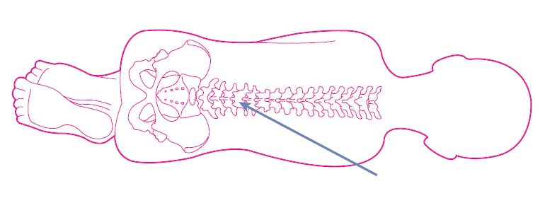
Children are asleep when this is performed and therefore feel no pain. However, in view of pressure changes in the CSF, it is advised to remain lying down for at least 2 hours to avoid nausea and headaches.
The clean and dry dressing must remain in place for two days after the procedure.
.
Bone marrow aspiration
Objective
The bone marrow aspiration is performed in the bone of the pelvis. This allows you to remove a small amount of bone marrow. The bone marrow is where blood is produced: red bloods cells, white blood cells and platelets. This examination is used to see whether their production is impeded by the presence of abnormal cells that are taking up all the space. This is called a myelogram.
In practice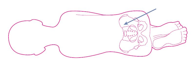
Children are asleep when this is performed and therefore feel no pain. When they wake up, they may have pain comparable to that of a bruise. Do not hesitate to ask for pain relief.
The clean and dry dressing must remain in place for two days after the procedure.
Standard X-ray
The X-rays used to perform an X-ray go through the tissues of the body differently according to their density (bone is harder than fat for example). This technique is used to obtain an overall image of the area being X-rayed. This is a painless and quick examination.
CT scan
This is a painless technique that obtains images of “slices” of the body. The X-rays are the same as those used for radiographic images. Sometimes, an iodine-based product is injected during the examination to better distinguish between healthy tissue and cancerous tissue. The duration of the examination is variable and rarely exceeds thirty minutes.
Scintigraphy
Like the PET Scan, this examination involves the injection of an isotope and is used to examine areas that consume a lot of energy in the body. This examination is particularly indicated for monitoring bone tumors.