Treatments in children
Based on the results of the examinations, age of the child and their response to initial treatments, a treatment protocol is chosen. The chosen protocol has, most of the time, been developed at international level. Indeed, as childhood cancers remain a rare group of diseases in our small country, international consensuses guide doctors in the choice of treatments for your child.
This protocol specifies the frequency, intervals, doses and administration duration of the various different medications. The duration, number of medications received or even the intensity of side effects are not directly related to the prognosis of the disease. We should therefore not compare the treatments between different children, but instead discus them with the doctor to understand the importance and role of each stage of treatment.
Chemotherapy
Chemotherapy drugs are medications that kill cancer cells throughout the whole body. There are many different chemotherapy drugs. Several different chemotherapy drugs are often combined to increase the efficacy of the treatment.
Sometimes they are used as the first line treatment and sometimes they are used as adjuvant to other treatments: surgery or radiotherapy for example.
Generally-speaking, chemotherapy drugs are always administered in several cycles to give the child’s healthy cells time to “recover” in between two cycles and to ensure that all of the persistent cancer cells are destroyed. By analogy with a garden, the first cycles pull out all the weeds and the subsequent cycles kill the roots.
Most chemotherapy drugs attack cells that reproduce rapidly such as cancerous cells which divide more quickly than their normal counterparts.
Routes of administration
Chemotherapy is administered either during a stay in hospital, in a day unit, or at home. Chemotherapy can be administered via various different routes: intravenous, intramuscular, subcutaneous or by mouth.
Most chemotherapy drugs irritate the small veins and must be administered via a large blood vessel. Different techniques are used to directly inject the drug into the blood vessel.
The central route
A long catheter is inserted through a vein in the neck or under the collarbone by the anesthetist. This procedure is carried out under light anesthetic. In contrast to the port, the central route is removed when the patient is discharged from hospital.
Implanted Port (Port-A-Cath®)
This is a small disc attached to a catheter which is inserted into a large blood vessel. This disc, which looks like the end of a stethoscope, is inserted under general anesthetic by the surgeon and placed just under the skin of the upper chest and sutured to the muscle. It has a membrane which allows blood to be taken and medications to be administered.
To make puncturing the port less painful, it is advised numb the child’s skin over the port with Emla® (anesthetic cream) one hour before drip is set up. After the port has been used, the dressing applied by the nurses should be kept dry and clean for 48 hours.
If your child has to come back several days in a row and the port needle could not stay in place, a new tube of Emla should be used before each infusion to avoid the risk of infection.
If the period of time between two consultations is spaced out, the port should be flushed approximately every 3 months to ensure it works properly.
The port is removed at the end of treatment after discussion with the doctors.
Radiotherapy
Radiotherapy uses ionizing radiation to destroy rapidly multiplying cancerous cells. This technique requires a number of calculations to ensure that the radiation beam only targets the cancerous cells and spares the health tissue around the tumor.
Before the first radiotherapy session, a “simulation” is organized in order to define the area to irradiate as accurately as possible.
Patients will then receive a timetable of sessions typically spread over a few weeks. The procedure does not often take more than a few minutes. However, during the session it is essential that the child remains completely still.
For more information, please consult this link, concerning the Vladi project.
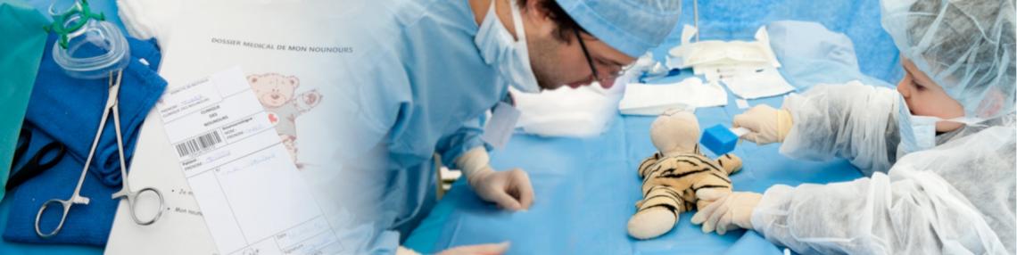
Surgery
Faced with tumors, surgery is often of great importance. In certain situations, it is able to remove most of the tumor. However, the majority of the time, even in the case of full surgical excision with the naked eye, treatment needs to be supplemented with radiotherapy or chemotherapy to ensure that the “roots” of the tumor have been destroyed.
Surgery is also very useful for the diagnosis of a mass by means of a biopsy: a small fragment of the tumor is sampled and examined under a microscope. For some tumors, other examinations performed on the fragments are used to determine the aggressiveness of the tumor and its response to different therapies.
La greffe de moelle osseuse
Dans certaines pathologies, un traitement par greffe de cellules souche provenant de la moelle osseuse peut être nécessaire. Ces cellules souches ont pour rôle de fabriquer les cellules qui composent le sang : les globules rouges, les globules blancs et les plaquettes.
Elles proviennent soit d’un prélèvement médullaire (cellules souches médullaires), soit d’un prélèvement périphérique (cellules souches périphériques), soit de sang placentaire (cellules souches de sang de cordon).
Selon l’indication de la greffe, ces cellules proviennent soit de l’enfant lui-même (autogreffe), soit d’une tierce personne compatible : un frère ou une sœur, un donneur volontaire anonyme (allogreffe) ou un sang de cordon disponible dans une banque de sang de cordon.
L’autogreffe est indiquée suite à des traitements intensifs qui ont détruit les cellules souches afin de reconstituer plus rapidement la moelle osseuse de l’enfant. Elle n’est en général pas utilisée pour les traitements des leucémies et des lymphomes.
Par contre, l’allogreffe est entre autres indiquée dans le cas de leucémies et de lymphomes. Le but de la greffe est de remplacer les cellules souches malades du receveur par celle du donneur compatible.
Pour ce faire, on administre à l’enfant une chimiothérapie ou une radiothérapie ayant pour but de détruire toutes les cellules cancéreuses. Ensuite, la poche contenant les cellules souches du donneur est administrée comme une simple perfusion afin de recoloniser la moelle du receveur.
La période post-greffe durant laquelle la moelle du receveur est détruite et la nouvelle moelle pas encore productive est très délicate. En effet, l’usine qui fabrique le sang est à l’arrêt complet durant 2 à 3 semaines en moyenne. En particulier, l’enfant est dépourvu de défense contre les infections. C’est pourquoi ces greffes ont lieu dans l’unité 56, l’unité stérile. La récupération médullaire est progressive et justifie une hospitalisation d’une durée totale de minimum 4 à 5 semaines. Ensuite, un suivi médical très rapproché avec de fréquentes perfusions d’immunoglobulines sera instauré.